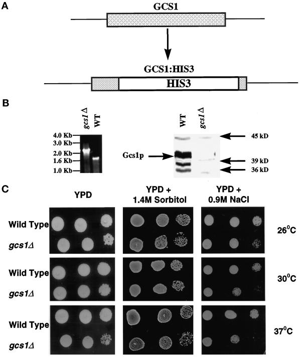
Figure 3.
Disruption of GCS1. (A) GCS1 was disrupted by replacing nucleotides 224-1021 with the HIS3 gene by homologous recombination. (B) Genomic DNA isolated from wild-type and gcs1Δ was screened by PCR (left). Whole-cell lysates were prepared from midlogarithmic wild-type and gcs1Δ cells grown at 30°C were separated by SDS-PAGE and transferred to nitrocellulose. Gcs1p was detected by immunoblot analysis using antisera generated against full-length His6-Gcs1p fusion protein (right). (C) Serially diluted cell suspensions (10 μl) were spotted onto YPD and YPD plates supplemented with either 1.4 M sorbitol or 0.9 M NaCl. The plates were incubated for 48–72 h at the indicated temperatures.

