Figure 7.
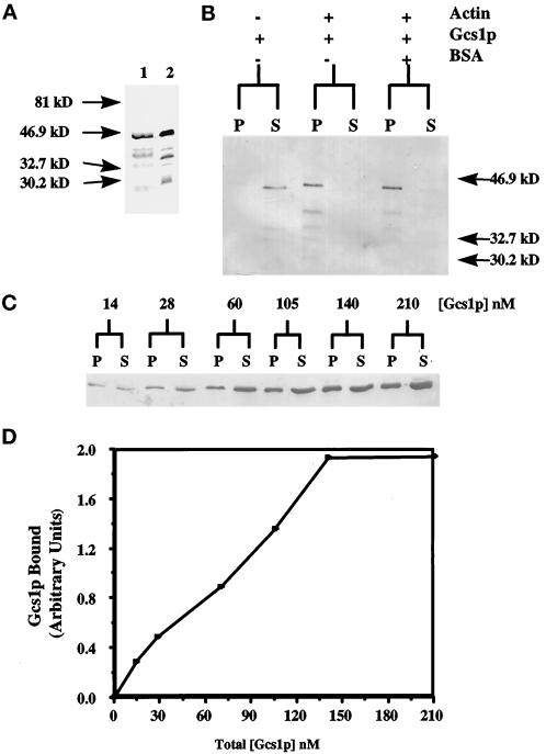
Gcs1p interacts with actin in vitro. (A) His6-Gcs1p was purified using Ni2+-NTA resin. The purified protein was analyzed by Coomassie stain (lane 1) and by immunoblot analysis using an anti-RGS-His4 antibody (lane 2). (B) Purified His6-Gcs1p was incubated with 3 μM polymerized yeast F-actin in the presence or absence of BSA and centrifuged at 270,000 × g. The pellets (P) and supernatants (S) were separated by SDS-PAGE and subjected to immunoblot analysis using an anti-RGS-His4 antibody. (C) Increasing concentrations (14–210 nM) of purified His6-Gcs1p were incubated with 3 μM polymerized yeast F-actin and centrifuged at 270,000 × g. The pellets (P) and supernatants (S) were separated by SDS-PAGE and subjected to immunoblot analysis using an anti-RGS-His4 antibody. (D) Quantification of immunoblot in panel C using a densitometer.

