Figure 8.
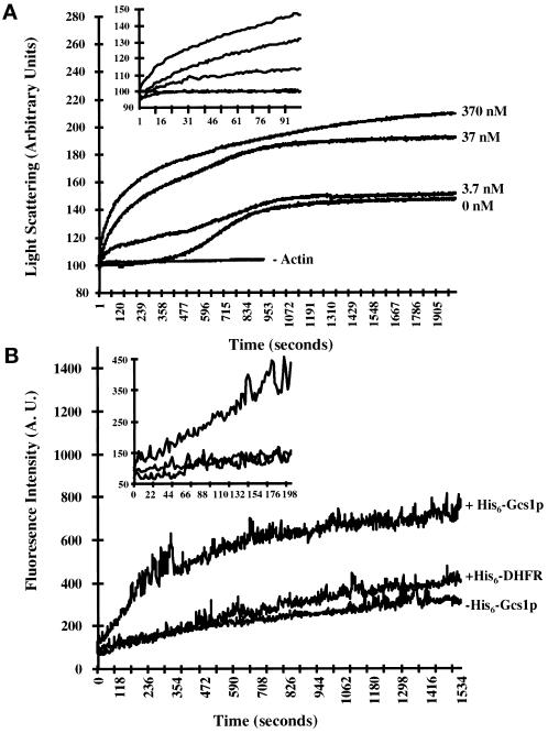
Time course for actin polymerization in the presence of His6-Gcs1p. (A) Monomeric yeast G-actin (2 μM) was polymerized in the absence or presence of increasing concentrations of His6-Gcs1p. Actin polymerization was monitored as an increase in light scattering as described in MATERIALS AND METHODS. Inset trace is a magnification of the first 105 s of the polymerization assay. In addition, 370 nM His6-Gcs1p was incubated in the absence of actin to determine the intrinsic light scattering of His6-Gcs1p. (B) Monomeric 1:10 pyrene-labeled rabbit muscle actin (5 μM): muscle actin was polymerized in the absence or presence of 0.2 μM His6-Gcs1p or His6-DHFR. Polymerization was monitored as an increase in fluorescence intensity as described in MATERIALS AND METHODS. Inset trace is a magnification of the first 300 s of the polymerization assay.

