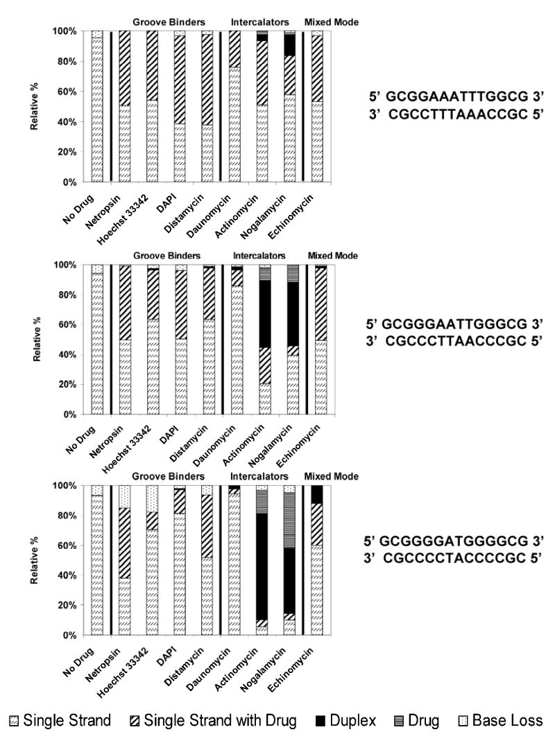
Figure 7.
IRMPD MS/MS (50 W, 0.8 ms – 1.6 ms) comparison for the [M - 6H]6− complexes containing one of three duplexes with varying GC character (sequences shown to the right of the bar graph) and one drug, grouped by their different modes of binding. Fragment ions were grouped by type including drug-free single strand ions, single strand ions with drug bound, drug-free duplex ions, free drug ions, and base loss ions. The bar graphs show the percentage for each of these fragment types with the total fragment ion current normalized to 100%.

