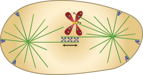
Fig. 2.
The mitotic spindle. Microtubules (green) form two asters and the central spindle, where the microtubules growing from the two spindle poles meet. Microtubule “plus-ends” (the more dynamic ends) are in the center of the spindle and at the cell periphery, while the “minus-ends” (the less dynamic ends) are anchored at the spindle poles. Motor proteins (blue, at the cell center) cross-link the overlapping anti-parallel microtubules in the central spindle and slide them past each other. Chromosomes (red; only one is shown for clarity) are attached to microtubules at the kinetochore (yellow), where forces towards and away from the spindle pole are generated. Chromokinesins (grey) reside at the chromosome arms and push the chromosomes away from the spindle pole by walking towards the microtubule plus-end. Minus-end directed motors (blue, at the cell periphery) are anchored at the cell cortex and pull on astral microtubules, thereby elongating the spindle

