
Figure 2.
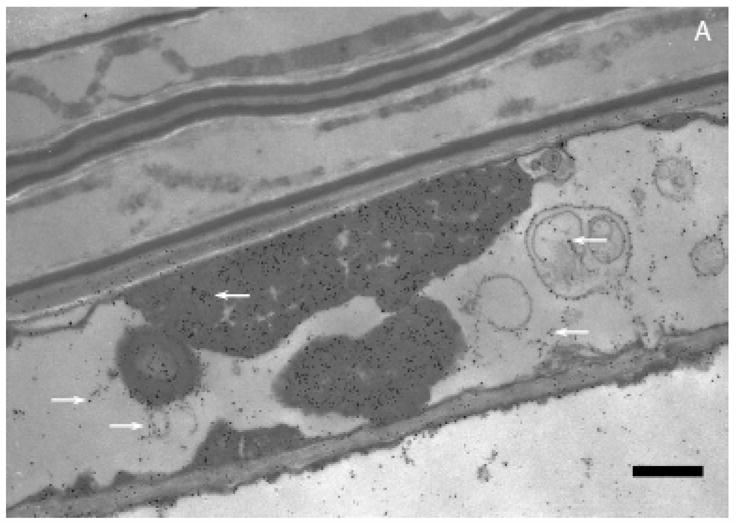
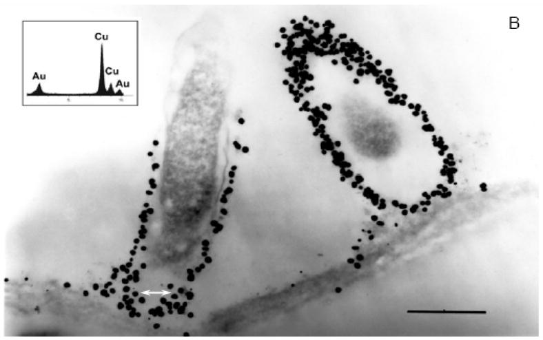
A. Transmission electron microscopy of Sesbania drummondii root cells loaded with gold nanoparticles (magnification = 20,000X; scale marker = 1.0 μm). B. Transmission electron microscopy of Sesbania drummondii root showing numerous gold nanoparticles surrounding organelles (magnification = 60,000X; scale marker = 500 nm). The inset on the top left shows energy-dispersive spectroscopic spectrum with gold and copper peaks (copper peaks arise from the copper grid that holds the plant tissue).