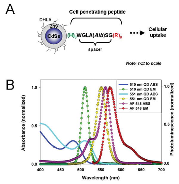
Figure 1. Schematic representation of a QD-peptide assembly and relevant absorbance/emission spectra for materials used in this study.

(A) The peptide self-assembles onto the DHLA-capped CdSe/ZnS QD surface via (His)8-Zn metal-affinity coordination. A short spacer separates the (His)8 QD attachment domain from the HIV-1 Tat-derived (Arg)8 cellular uptake sequence. Aib is α-amino isobutyric. (B) Absorbance of 510 nm and 551 nm QDs are represented by the solid black and cyan lines, respectively. The fluorescence spectra of the QDs and AlexaFluor 546 are also shown.
