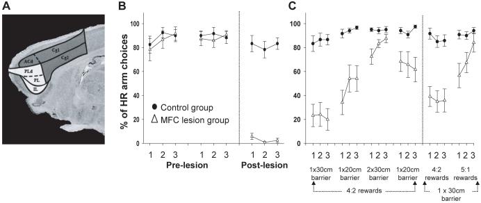
Figure 4.
(A) Representation of the targeted MFC (all shaded areas) and ACC regions (dark gray shading only). In both experiments reported here, lesions were complete and restricted to the targeted cortical regions as intended (see Walton et al., 2002, 2003 for detailed histology). (B) Performance of groups of control and MFC-lesioned rats on a T-maze cost-benefit task both before and after excitotoxic lesions. Animals chose between climbing a 30 cm barrier to achieve a larger reward (HR) or selecting to obtain a smaller quantity of food from the vacant arm. (C) Choice behavior of control and MFC-lesioned rats postoperatively in response to changes in the effort quotients in each goal arm (as denoted by the size of the barriers) or the reward ratio between the HR and LR.

