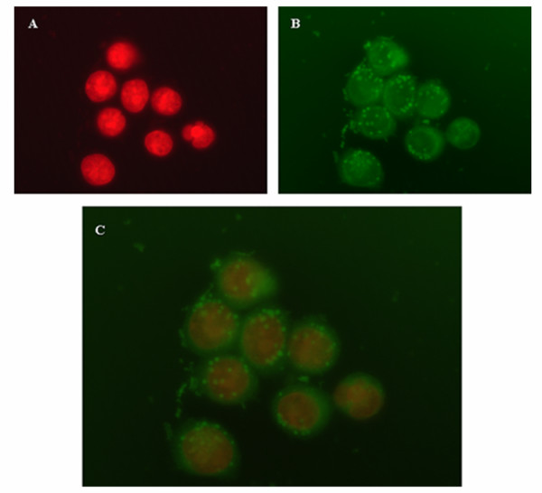
Figure 3.

Intracellular distribution of NS in ARO cells. ARO cells were incubated for 2 h with PLGA NS loaded with the fluorescent coumarin-6 probe and analyzed by fluorescent microscopy. Nuclei were stained with PI and are visible in red (1A). The uptake of coumarin-6-loaded NS is visible in green (1B). Figure 1C displays an overlaying images obtained combining the FITC and the PI filters. A representation of two experiments is shown. Magnification: 63 ×
