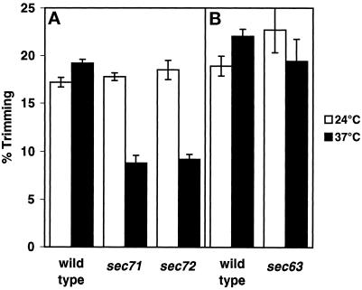
Figure 3.
sec71Δ and sec72Δ, but not sec63-1, membranes show a defect in the in vitro ER–nuclear membrane fusion assay at 37°C. (A) Donor and acceptor membranes (75 μg protein each) prepared from wild-type strains (MLY1601 and MLY1600), strains deleted for the SEC71 gene (MLY1889 and MLY1890), or strains deleted for the SEC72 gene (MLY1891 and MLY1892) were combined in the presence of an ATP regeneration system in a total volume of 50 μl and held on ice. Reactions were incubated for 60 min at 24 or 37°C. (B) In a separate experiment, donor and acceptor membranes prepared from wild-type strains MLY1600 and MLY1601 and sec63-1 strains MLY1651 and MLY1652) were combined and incubated as above. In all cases, the experiments were repeated three times, and the mean values and SDs are shown. All strains were grown at 24°C before membrane isolation. The amount of glucose trimming, indicative of the successful fusion of membranes, was assessed as described previously (Latterich and Schekman, 1994).

