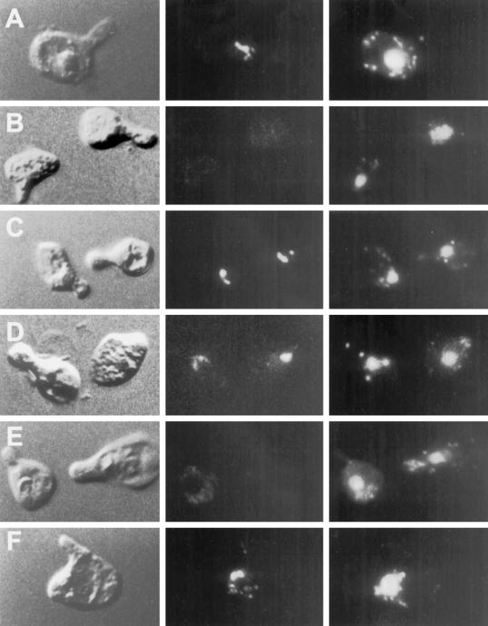
Figure 4.
Kar5p is mislocalized in kar7-1039 but not in other karyogamy mutants. In each series (A–F), the left panel shows the morphology of the cell (shmoo) by DIC. The middle panel shows the Kar5p immunofluorescence, and the right panel shows the nucleus stained with DAPI. (A) Kar5 shmoos (MS3987); (B) kar5Δ2 shmoos (MS3986); (C) kar1-1 shmoos (MS4021); (D) kar2-1 shmoos (MS4020); (E) kar7-1039 shmoos (MS3991); (F) kar8-1333 shmoos (MS3989). Cells were treated with α-factor for 2–2.5 hr before preparation for immunofluorescence. A and B are reproduced from Beh et al. (1997) J. Cell. Biol. 139, 1063–1076, by copyright permission of The Rockefeller University Press.

