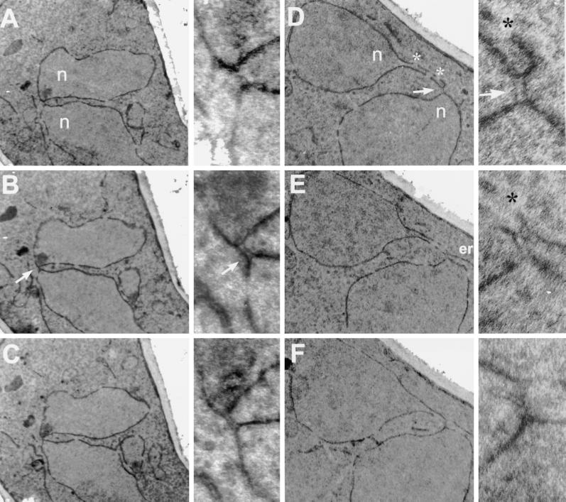
Figure 6.
Electron micrographs of serial section of two kar8-1333 zygotes. The kar8-1333 mating partners used in this study were MS2705 and MS2706. (A–C) Micrographs of three consecutive serial sections (of seven) through a kar8-1333 mutant zygote shown at two different magnifications. (D–F) Micrographs of sections 1, 3, and 5 (of five) of another kar8-1333 mutant zygote also shown at two different magnifications. Each section is 70 nm thick. n, nuclei; *, nuclear pores; arrows, kar8-1333 bridges between the two nuclei.

