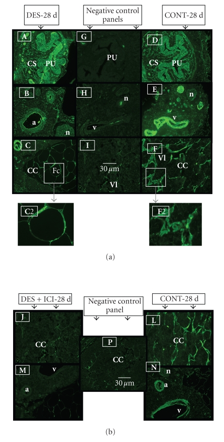
Figure 6.
(a) Representative immunohistochemical staining for PPARγ protein in the body of the penis of 28-day-old DES-treated (A), (B), and (C) and control untreated rats (D), (E), and (F). Note that PPARγ protein is expressed in DES-treated (DES-28 d) and normal rats (CONT-28 d) with increased intensity and fat cells in DES-treated rats (see panel (C)). Note expression in transitional epithelium of the penile urethra (PU) and the surrounding corpus spongiousm (CS) in (A) and (D) and in the endothelium of blood vessels and smooth muscle cells in the dorsal artery (a) and vein (v), and in nerve fibers of the dorsal nerve (n) of the penis (B) and (E). Similar staining intensity can be seen in the endothelium and smooth muscles of the vascular lacunae (Vl) in the corpus cavernosum penis (CC) in control normal rats (F). Note one contrasting difference is that the cavernous spaces in DES-treated rats in panel (C) are replaced with fat cells (Fc) that show increased staining intensity in the cell nucleus located at cell periphery. Panels (C2) and (F2) show a closer view of area outlined by insert box. Control sections (minus primary antibody) were in panels (G), (H), and (I). Scale bar = 30 μm. (b) IHC staining for PPARγ protein was significantly reduced by ICI 182, 780 treatment [compare staining in panels (J) and (M) with (L) and (N)]. Panel (P) is a negative control (minus primary antibody). Scale bar 30 μm.

