Fig. 4.
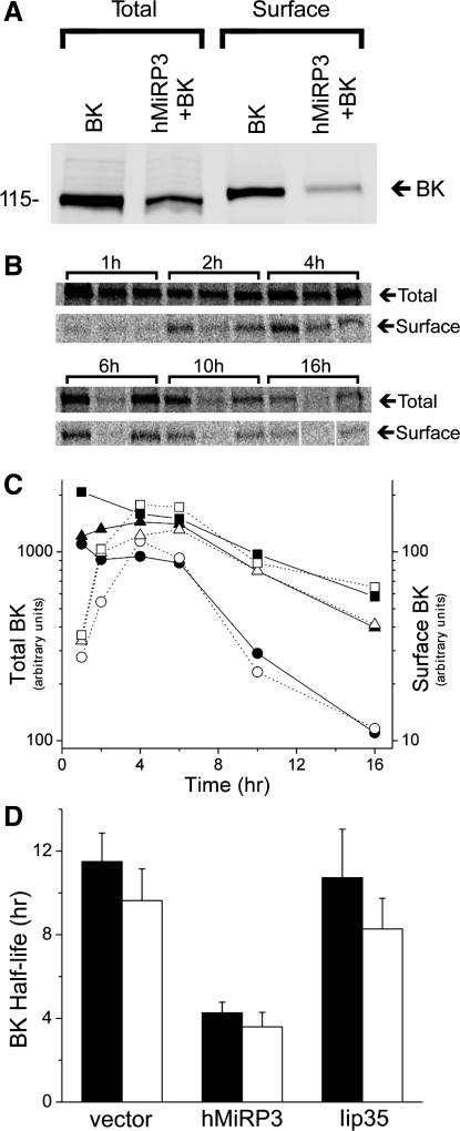
MiRP3 shortens half-life of BK protein. A: CHO cells were transfected with wild-type BK channels and an equal concentration of either empty vector (BK) or MiRP3 (hMiRP3+BK), allowing quantification of the total and surface-expressed BK. The sample blot shows cotransfection of MiRP3 with BK leading to a 61% reduction of total BK and an 84% reduction of surface BK at steady state. B–D: pulse-chase experiments demonstrating enhanced degradation of cellular BK by MiRP3. B: phosphorimages of total cellular BK and surface-expressed BK chased for the times specified above the brackets. For each time point, the samples were loaded–from left to right–from cells expressing BK with empty vector, MiRP3, and Iip35. C: densities of the bands in B were quantified for the 16-h period (empty vector, squares; MiRP3, circles; Iip35, triangles). Arbitrary counts for total cellular expression (filled symbols) and surface expression (open symbols) are plotted to the left and right y-axes, respectively. D: half-life of BK expression for 3 experiments is plotted for total cellular expression (filled bars) and surface expression (open bars, means ± SE).

