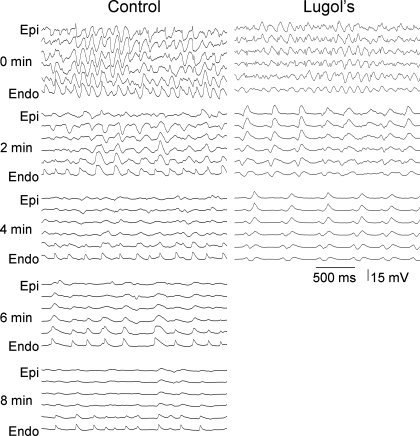
Fig. 4.
Recordings from the 6 electrodes of a plunge needle in a control and a Lugol-ablated heart every 2 min during VF. As VF progresses, an activation rate gradient develops in the control animals in which the endocardium (Endo) activates more rapidly than the epicardium (Epi). In the Lugol-ablated heart, the endocardium does not activate more rapidly than the epicardium. VF terminated at 8.25 min in the control heart and at 4.5 min in the Lugol-treated heart shown in this figure.

