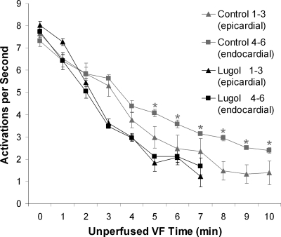
Fig. 5.
Mean activation rate of the 3 most endocardial and the 3 most epicardial plunge needle electrodes during VF. An activation rate gradient developed after 4 min of VF in the control hearts with the endocardial electrodes activating significantly faster than the epicardial electrodes but did not develop in the Lugol-ablated hearts. Mean activation rate with the standard deviation is shown. Statistically different values are denoted with asterisks.

