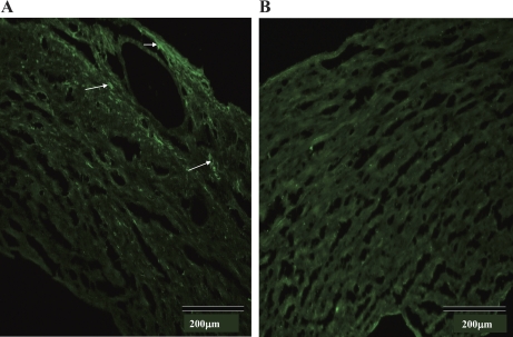
Fig. 8.
Immunofluorescence labeling of TRPV1 in WT (A) and TRPV1−/− (B) hearts. TRPV1 positive staining as indicated by the arrows is found in the myocardium and vessels in the left ventricle of the WT heart (A) but not the TRPV−/− heart (B). Negative staining was also found in WT control hearts in which the primary antibody was omitted from the immunofluorescence assay (data not shown).

