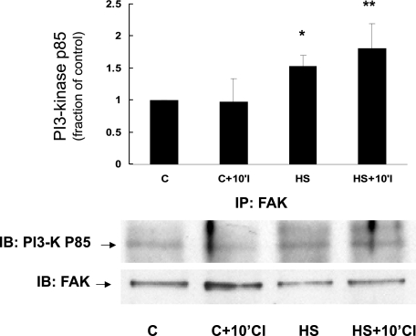
Fig. 3.
Effect of HS on interaction between FAK and PI3K in NRVM. Cell lysates from control myocytes or myocytes subjected to HS were prepared and immunoprecipitated (IP) with anti-FAK antibody and then subjected to electrophoresis. The resulting Western blot was stained with anti-PI3K-p85 antibody (IB; bottom). Compared with control myocytes, there was increased interaction between FAK and PI3K in myocytes subjected to HS, consistent with assembly and/or increased interaction between 2 key members of the proposed signaling complex (*P < 0.029, C vs. HS, n = 4; **P < 0.06, C+10'CI vs. HS+10'CI, n = 4). y-Axis data are plotted as fraction of control myocyte levels. The total amount of FAK protein was not significantly different between the various groups.

