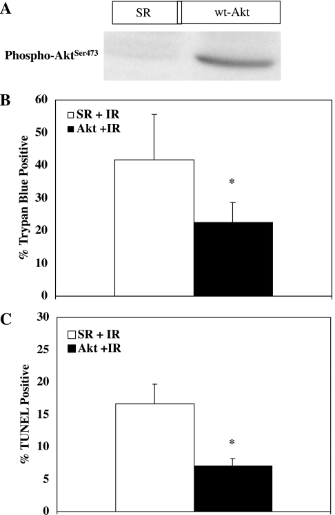
Fig. 5.
Effect of AKT on lethal cell injury in cultured myocytes. A: NRVM were infected with an adenovirus (adv) designed to increase expression of AKT as described in materials and methods. A Western blot demonstrating the effect of adv-AKT on AKT phosphorylation is shown. Compared with myocytes infected with an empty adenovirus (SR), myocytes infected with wild-type (wt) adv-AKT contained significantly more activated (phosphorylated) AKT. B: NRVM were infected with either empty virus or virus designed to increase expression of AKT and then subjected to 30 min of simulated ischemia followed by 30 min of reperfusion (IR). y-Axis values indicate % of cells staining positive with Trypan blue (dead cells). Increased expression of AKT resulted in significant protection against oncotic cell death. *Significant difference from control cells (P ≤ 0.02; n = 5). C: NRVM were infected with either empty virus or virus designed to increase expression of wild-type AKT and then subjected to 30 min of simulated ischemia followed by 30 min of reperfusion. y-Axis values indicate % of TUNEL-positive cells (apoptotic cells). Increased expression of AKT resulted in significant protection against apoptosis. *Significant difference from control cells (P < 0.005; n = 5).

