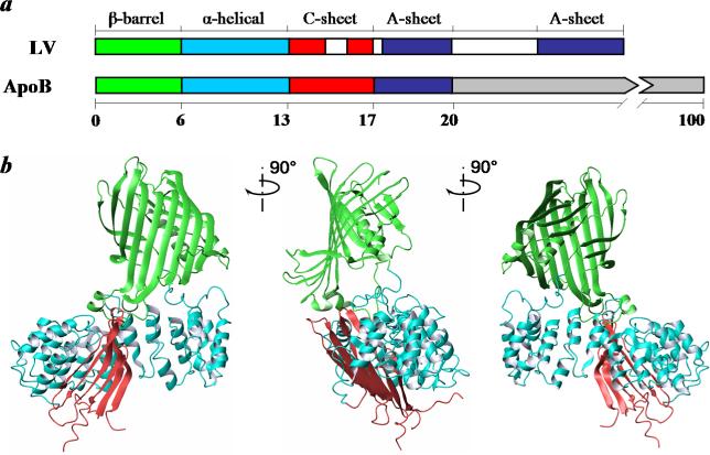
Figure 1. Proposed domain organization in B17.
a. The domain comparison between lipovitellin (LV) and apoB. The homology between LV and apoB extends to approximately B20. The five domains in LV are colored by β-barrel, green; α-helical, cyan; C-sheet, red; A-sheet, dark blue (12). Regions missing in the crystal structure of LV are in white. ApoB sequences not homologous to LV are shown in gray. b. The three dimensional model of B17 based on LV. The three domains in B17 are colored in the same scheme.

