
Figure 2. AktThr-308 and AktSer-473 kinase activities in Epac1 immunoprecipitate of plasma membrane and nuclei of macrophages.
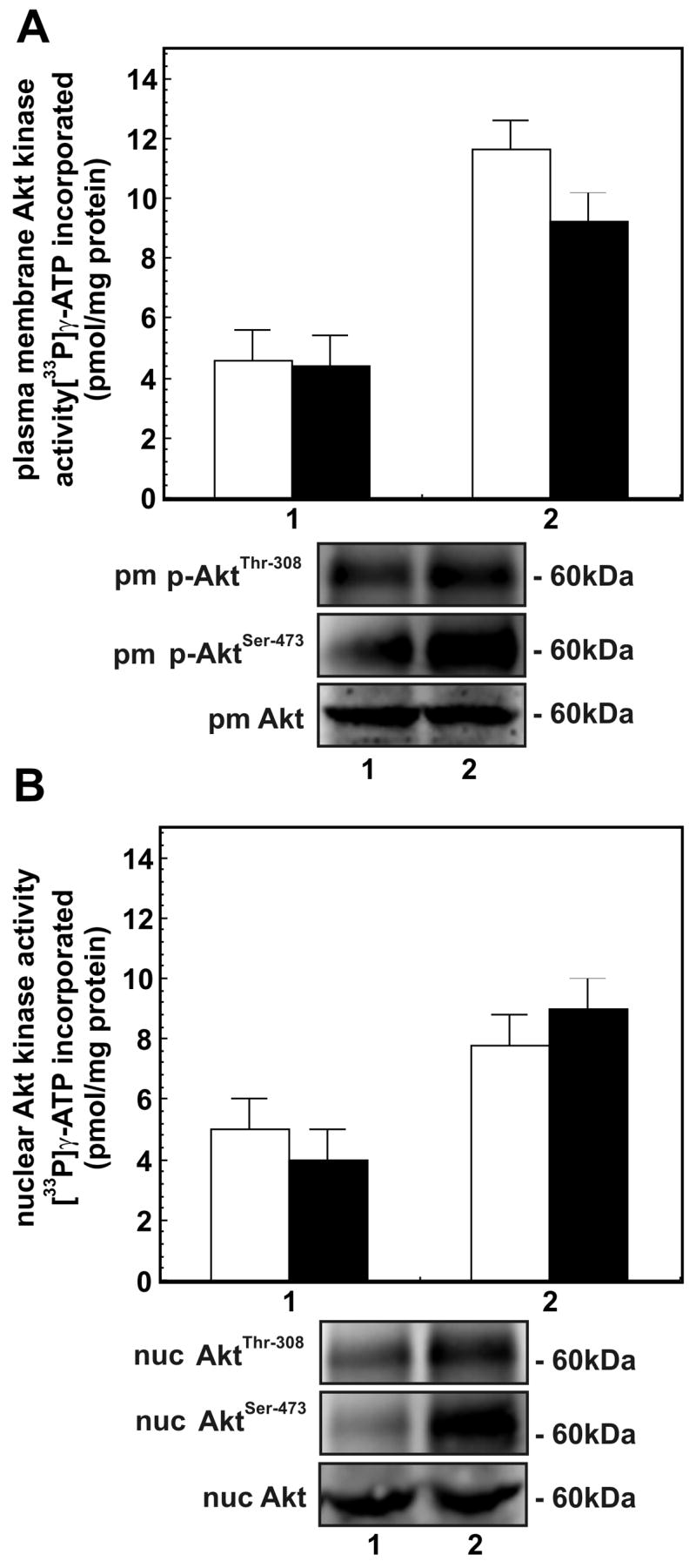
Panel A - AktThr-308 and AktSer-473 kinase activities in plasma membranes of macrophages treated with: (1) buffer; and (2) 8-CPT-2-O-Me-cAMP (200 μM/30 min). The bars are: [□] AktThr-308 kinase activity and [■] AktSer-473 kinase activity. Panel B - AktThr-308 and AktSer-473 kinase activites in nuclei of cells treated as in Panel A. The bars are: [□] AktThr-308 kinase activity and [■] AktSer-473 kinase activity. Both Akt kinases activities in Epac1 immunoprecipitates of plasma membranes and nuclei are expressed as pmol [33P]γ-ATP incorporated into respective peptides/mg protein and are mean ± SE from three independent experiments. The immunoblots showing changes in levels of p-AtkThr-308 and p-AktSer-473 in plasma membranes and nuclei of cells treated as in Panel A are shown below respective panels. Immunoblot of protein loading control Akt is also shown.