
Figure 5. Subcellular localization of Epac1 p-Akt1Thr-308 and p-AktSer-473 in cells treated with 8-CPT-2-O-Me-cAMP as determined by confocal microscopy.
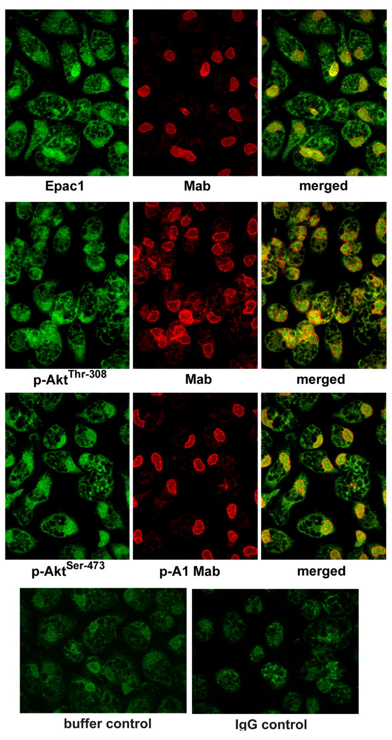
See “Experimental Procedures” for details. The localization of Epac1, p-AktThr-308, and p-AktSer-473 (green) in the perinuclear nuclear region stimulated cells compared to unstimulated cells was determined using a nuclear marker Mab414 (red). Perinuclear localization of Epac1, p-AktThr-308, and p-AktSer-473 in stimulated cells is seen on merging of green and red fluorescence. The results shown are representative of six independent experiments.