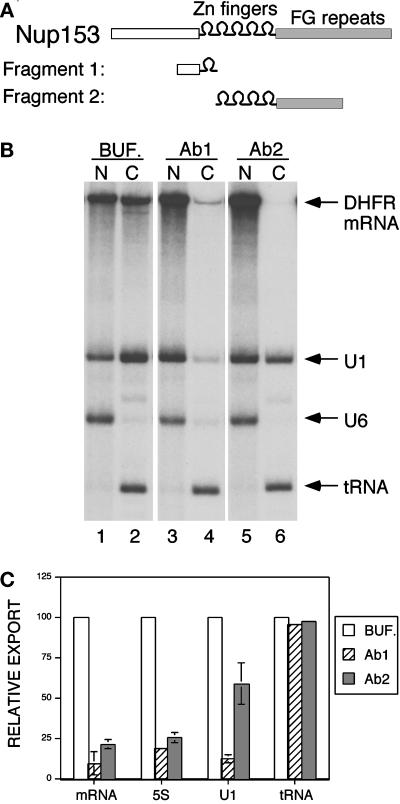
Figure 6.
Antibodies to distinct regions of Nup153 affect export differentially. (A) The domain structure of Xenopus Nup153 is represented schematically, showing the unique N-terminal region, the five zinc fingers, and the FG-containing C-terminal region. Antibody 1 (Ab1) was raised against a fragment containing part of the N-terminal region and one zinc finger (amino acids 53–334). Antibody 2 (Ab2) was raised against the remaining four zinc fingers and part of the FG region (amino acids 334–828). (B) Oocytes were injected as described in Figure 1, first with buffer or antibodies and then with radiolabeled RNA. The RNA mixture used here contained DHFR mRNA (an intronless mRNA), U1 snRNA, U6 snRNA, and tRNA. RNA localization was analyzed after a 4 h incubation. DHFR mRNA export was inhibited equally by Ab1 and Ab2, whereas U1 RNA export is significantly less sensitive to Ab2 (compare lanes 2, 4, and 6). (C) The effects of Ab1 and Ab2 are depicted in this bar chart in which data from multiple experiments are summarized. Inhibitory profiles of antibodies to distinct regions of Nup153 are shown here for the adenoviral mRNA (spliced), U1 snRNA, 5S rRNA, and tRNA.

