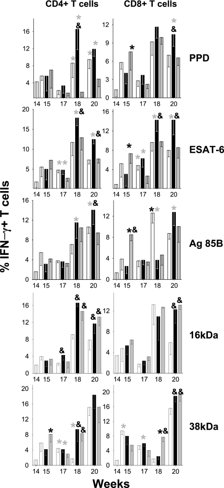
FIG. 4.
Evolution of CD4+ and CD8+ IFN-γ+-specific cells from spleen in aerosol-infected mice. After infection, mice were treated with INH/RPN from weeks 6 to 14 (in white) and with two subcutaneous inoculations of RUTI at weeks 14 and 17 (in black) or BCG at week 14 (in gray). Data are expressed as the percentages of means and SD. An asterisk or ampersand indicates significant differences (P < 0.05 by t test). The color of the asterisk corresponds to the group with lower values to which the comparison was significant. Black and gray asterisks refer to RUTI and BCG groups, respectively; the ampersand refers to the control group.

