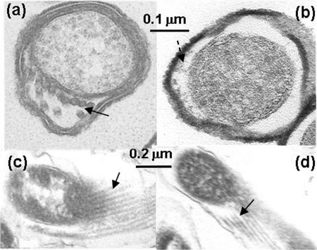
FIG. 4.
Electron microscopic analysis of the wild type and the flaB1 flaB2 mutant. (a and b) Thin section of the wild type and the flaB1 flaB2 mutant. The solid arrow points to PFs, and the dashed arrow points to amorphous staining material seen in the flaB1 flaB2 periplasmic space. (c and d) Oblique sections of the wild type and the flaB1 flaB2 mutant. Arrows point to PFs in panel c and to tubular material in panel d.

