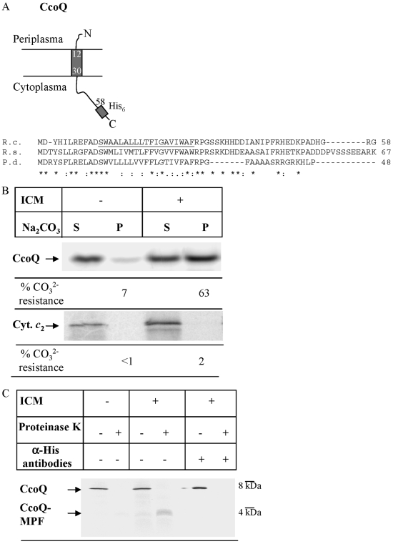
FIG. 1.
CcoQ is a type I integral membrane protein. (A) Predicted topology of CcoQ in R. capsulatus and sequence alignment of CcoQ from the closely related species R. capsulatus (R.c.), R. sphaeroides (R.s.), and P. denitrificans (P.d.). The alignment was performed by using CLUSTAL; an asterisk (*) in the sequence indicates identical amino acids, and a colon (:) indicates similar amino acids. The putative transmembrane domain of CcoQ in R. capsulatus is underlined. (B) CcoQ and cyt c2 were in vitro synthesized in the presence or absence of R. capsulatus inverted inner membrane vesicles (ICM) using a coupled R. capsulatus in vitro transcription-translation system. After in vitro synthesis the reaction mixture was extracted with alkaline Na2CO3, pH 11.3 and centrifuged. The supernatant (S) reflecting soluble proteins and the pellet (P) reflecting membrane-integral proteins were loaded on a 22% urea-SDS-PAGE (CcoQ) or on a 16.5% Tris-Tricine (cyt c2) gel. Radioactively labeled proteins were visualized by phosphorimaging. For quantification, the amount of soluble material and that of the pellet fraction was set as 100%, and the material present in the individual fractions was quantified. The quantification is based on at least three independent experiments. (C) For proteinase K protection, the in vitro reaction mixture was split, and one-half was directly precipitated for 30 min at 4°C with ice-cold TCA; the other half was incubated with proteinase K (0.5 mg/ml, final concentration) for 20 min at 25°C. The proteinase K-treated sample was also then TCA precipitated. After centrifugation of the TCA-precipitated samples, the pellets were resuspended in SDS-loading dye, heat denatured, and loaded onto a 22% urea-SDS-PAGE gel. Immunoprecipitation experiments were performed with α-His antibodies covalently bound to protein A-Sepharose beads. For proteinase K-treated samples, the protease inhibitor phenylmethylsulfonyl fluoride was added before the addition of the antibody beads.

