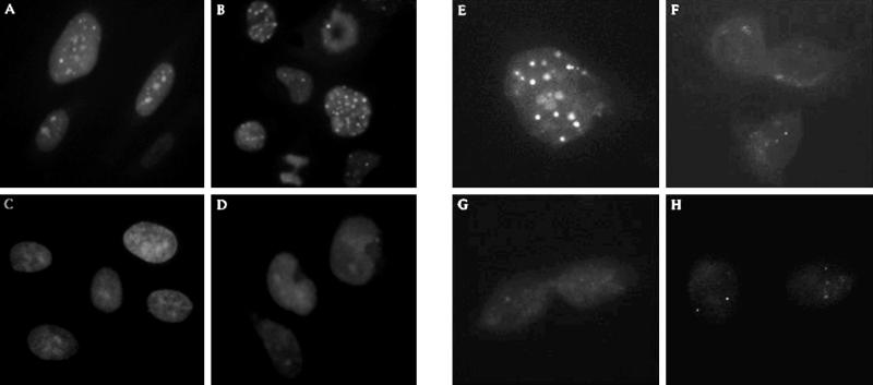
Figure 2.
Indirect immunofluorescence study of BLM in the nucleus of normal, Bloom syndrome, and SV40-transformed human fibroblast cell lines. All cells are fixed and stained with BLM antibodies, followed by donkey anti-rabbit secondary antibodies conjugated to Texas Red. Cells in A–E are stained with DAPI. Cells in F–H are not stained with DAPI to show diffuse BLM staining. (A) Normal human fibroblasts (HG2619). (B) SV40-transformed normal human fibroblasts (HG2855). (C) BS fibroblasts (HG2940). (D) SV40-transformed BS fibroblasts (HG2522). (E) HG2522 transfected with the normal BLM cDNA. (F) HG2522 transfected with the Q672R cDNA. (G) HG2522 transfected with the C1055S cDNA. (H) HG2522 transfected with the K695T cDNA.

