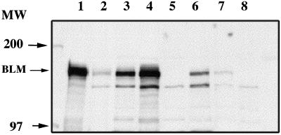
Figure 3.
Western transfer analysis of BLM proteins in cell lines and transfected clones. Twenty micrograms of total cell protein were loaded on each lane, and proteins were displayed on a 5% SDS polyacrylamide gel and transferred to a PVDF membrane. Positions of molecular weight markers and BLM are indicated by arrows. Lane 1: SV40-transformed normal fibroblast cell line (HG2855); lane 2: normal fibroblast cell line (HG2619); lane 3: SV40-transformed BS fibroblast cell line (HG2522) transfected with the normal BLM cDNA (R12c41); lane 4: HG2522 transfected with the normal BLM cDNA (R12c45); lane 5: HG2522; lane 6: HG2522 transfected with the Q672R allele; lane 7: HG2522 transfected with the C1055S allele; lane 8: HG2522 transfected with the K695T allele.

