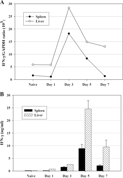
FIG. 2.
IFN-γ mRNA and IFN-γ ex vivo secretion are detected in both spleen and liver quickly after LVS infection. C57BL/6J mice were infected with 105 CFU of LVS i.d., and pooled single-cell suspensions of spleen (▪) and liver (□) cells were prepared from five mice at the indicated time points. (A) IFN-γ-specific mRNA prepared from splenocytes (▪) and liver leukocytes (□) at the indicated time points after LVS infection was quantified by real-time PCR and normalized in relationship to GAPDH expression. (B) To assess secretion of IFN-γ protein, splenocytes and liver leukocytes were cultured for 3 days, and the harvested supernatants were analyzed for secreted IFN-γ by ELISA. Results are means ± standard deviations for three individual cultures established using pooled cells as described above. Results shown were derived from a single representative experiment of two (panel A) or three (panel B) total experiments of similar design.

