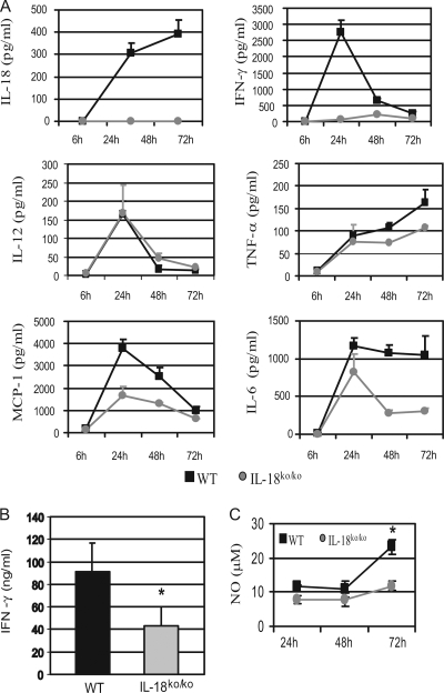
FIG. 3.
Cytokine pattern in IL-18ko/ko mice during the early phase of L. monocytogenes infection. (A) C57BL/6 and IL-18ko/ko mice were infected i.v. with 50,000 listeriae. Serum cytokine concentrations of these mice were assessed at the indicated time points after infection. The sera of four mice per group were pooled, and cytokine concentrations were determined by cytometric bead assay. (B) Three days after infection with 50,000 listeriae, 3 × 10⁶ splenic cells from infected mice were restimulated in vitro with 5 × 10⁷ HKL cells for 24 h, and the IFN-γ concentration in the supernatant was measured by enzyme-linked immunosorbent assay (*, P < 0.05). Cytokine levels in cultures which were not restimulated with HKL were below the detection limit. (C) After 24, 48, and 72 h of infection, splenic cells were restimulated with HKL for 24 h, and NO concentrations in the supernatants were determined as described in Materials and Methods. Data show one experiment representative of three performed.

