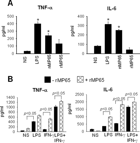
FIG. 6.
TNF-α and IL-6 production from activated MDM after stimulation with rMP65. (A) MDM were left untreated (NS) or treated with rMP65, nMP65 (5 μg/ml), or LPS (1 μg/ml) for 18 h at 37°C. After incubation, supernatants were recovered and tested for the presence of TNF-α and IL-6. Data are expressed as means ± SEM for four independent experiments. *, P < 0.05 (rMP65-treated cells versus untreated cells). (B) MDM were left untreated (NS) or treated with LPS (1 μg/ml) or IFN-γ (100 ng/ml), alone or in combination, for 30 min at 37°C. After activation, cells were left untreated (−rMP65) or treated with 5 μg/ml rMP65 (+rMP65) for 18 h at 37°C. Data are expressed as means ± SEM for four independent experiments. Statistical analysis was performed with ANOVA and corrected by the Bonferroni test for multiple comparisons. *, P < 0.05 (rMP65-treated cells versus untreated cells).

