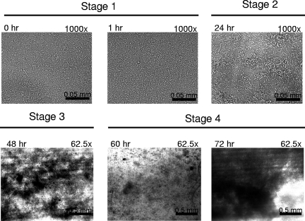
FIG. 1.
Bright-field microscopy of flow cells inoculated with L. monocytogenes 10403s. The flow cell at ambient temperature was inoculated and allowed to remain static for 1 h. Medium flow was then restored, and images of surface-attached cells were captured at a magnification of either ×1,000 to image individual surface-attached cells or ×62.5 to visualize masses of surface-attached cells (dark regions against light background). The time points and level of magnification for each image are indicted in the upper left and right, respectively. Stage 1, surface attachment of individual cells; stage 2, microcolony formation; stage 3, biofilm maturation; stage 4, community dissociation (60 h) followed by regeneration (72 h).

