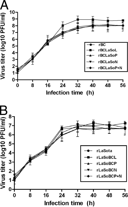
FIG. 2.
Multicycle growth kinetics of the parental and the chimeric NDVs in DF-1 cells. (A) BC viruses. (B) LaSota viruses. Six-well plates of DF-1 cell monolayers were infected with the virus at an MOI of 0.01 PFU per cell for 1 h. The cells were washed with phosphate-buffered saline and then overlaid with DMEM containing 5% FBS at 37°C in 5% CO2. Supernatant samples were collected at 8-h intervals until 56 h postinfection. The medium of all LaSota backbone viruses contained 10% fresh allantoic fluid. The six-well plates were replaced with equal volumes of fresh medium. Virus yields at different time points were determined by plaque assay. Error bars show standard deviations.

