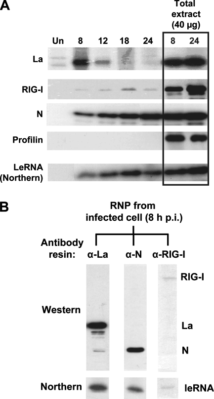
FIG. 2.
Protein composition of intracellular viral leRNP. (A) Western analysis of the total pool of infected cell RNP at various times postinfection. Equal amounts of uninfected (Un) or RSV-infected A549 cell extracts (at 8, 12, 18, and 24 h p.i.) were subjected to CsCl buoyant density centrifugation, and the materials from the RNP band region (i.e., fraction 4 in Fig. 1A) were subjected to Western analysis to detect La, RIG-I, or N. In parallel lanes (boxed), total cell extracts (equivalent to 40 μg protein) were directly subjected to Western analysis. Profilin serves as a negative control for leRNA binding and as evidence for equal loading in the total extract lanes. (Bottom) Northern analysis of the corresponding fractions to determine their leRNA contents. Note the high level of content of La in early-stage RNP, which is replaced by increasing N content of the RNP over time. (B) Protein-based selection and analysis of infected cell RNP. Equal amounts of CsCl-banded RSV-infected cell leRNP were incubated with protein A-Sepharose beads conjugated to antibodies against La, N, or RIG-I, as indicated above the lanes. Equal portions of the bound material from each column were analyzed by immunoblotting (Western) using a mixture of antibodies to La, N, and RIG-I. The remainder of the bound material was deproteinized, and the leRNA content was determined by Northern analysis (bottom).

