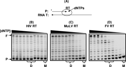
FIG. 1.
dNTP concentration-dependent reverse transcription activity of RT proteins. (A) Schematic illustration of primer extension reaction by RT. A 5′-end, 32P-labeled, 23-mer T primer (P, 5′-CCGAATTCCCGCTAGCAATATTC-3′) annealed the 38-mer RNA template (T, 5′-GCUUGGCUGCAGAAUAUUGCUAGCGGGAAUUCGGCGCG-3′; template/primer ratio, 2.5:1) was extended by RTs of HIV-1 (B), MuLV (C), and PFV (D), showing approximately 25, 60, and 75% of primer extension (F), respectively, with 250 μM dNTPs (first lane) at 37°C for 5 min as previously described (12), and the reactions were repeated with decreasing dNTP concentrations (125, 50, 25,10, 5, 1, 0.2, 0.1, and 0.05 μM). The dNTP concentrations found in dividing cells (D) (1∼5 μM) and macrophages (M) (0.05 μM) are marked at the bottom of the figure. All three RT proteins used here were fused to the N-terminal His tag and purified from a bacterial overexpression system as described previously (7). PFV RT was purified from pET28a, containing the PFV PR-RT gene provided by Stephen Hughes (1). F, 38-nucleotide-long, fully extended product; P, 23-mer, unextended primer.

