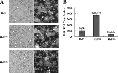
FIG. 7.
Syncytium formation analyses. (A) Formation of syncytia induced by HaF and HaF mutants expressed by infected cells. HzAM1 cells were infected with recombinant BVs (vHaBacΔF-HaFN1G, vHaBacΔF-HaFG3L, and vHaBacΔF-HaF) at an MOI of 5 TCID50 U/cell. Thirty-six hours after infection, cells were treated for 5 min with Grace's medium at pH 5.0. Syncytium formation was scored 24 h after exposure to acid conditions by phase-contrast microscopy, light (left) and fluorescence (right) micrographs. (B) Quantification of the ability of parental HaF protein and mutant HaF proteins to form syncytia. Each column represents the percentage of parental HaF fusion, and the data are derived from triplicate infection experiments. The error bars represent standard deviations from the means. For details, see Materials and Methods.

