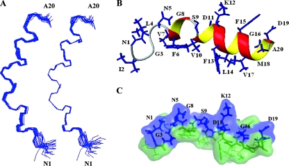
FIG. 9.
Structure of the fusion peptide in SDS micelles at pH 5, 27°C by 1H-NMR. (A) Superposition of the backbone atoms of the best 20 structures. The structures are fitted to residues 1 to 20 (left) and residues 7 to 18 (right). (B) Ribbon representation of the structure closest to the mean, with the side chains shown as blue sticks and indicated by residue name and number. (C) Surface representation of the structure closest to the mean, with hydrophilic surface in blue and hydrophobic surface in green. The side chains of hydrophilic residues are shown as blue sticks and indicated by residue name and number.

