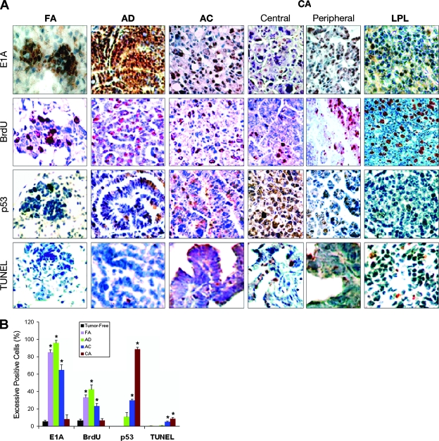
FIG. 4.
High levels of E1A 243-aa protein lead to high levels of proliferation rather than high levels of p53 and apoptosis. (A) Representative lung sections of focal adenomas (FA), adenomas (AD), adenocarcinomas (AC), and central and peripheral areas of carcinomas (CA) from E1A+E1B transgenic mice and LPL from A97 E1A transgenic mice stained for E1A 243-aa protein, BrdU-incorporating proliferation, p53, and TUNEL-positive apoptosis. Excessively stained E1A protein-positive nuclei (brown) or BrdU-positive nuclei (red) presented in tumor cells. BrdU-positive nuclei also existed in the proliferating lymphocytes of LPL. Excessively p53-positive nuclei (brown) mainly appeared in the tumor cells of AC or the CA central area. Few tumor cells in AC or CA showed TUNEL-positive nuclei (red). Original magnifications: ×100 (FA) and ×40 (AD, AC, CA, and LPL). The percentages of excessively stained E1A and p53-, BrdU-, and TUNEL-positive cells in tumor-free lung and FA, AD, AC, and CA central area of E1A+E1B transgenic mouse lungs are dynamically shown in panel B. Columns, means (n = 3); error bars, standard deviations; *, P < 0.025 versus tumor-free lungs.

