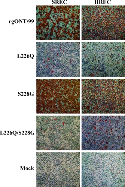
FIG. 1.
Infectivity levels of the H4N6 influenza viruses in SRECs and HRECs. Cells were infected with three TCID50s/cell for each virus, and infected cells were identified by immunocytochemistry (ICC) using the anti-influenza A NP antibody 68D2 (kindly provided by Y. Kawaoka, University of Wisconsin-Madison). Following ICC staining, the brightness and contrast of HREC micrographs were adjusted with ACDSee Photo Editor (ACD Systems) and PowerPoint (Microsoft) software to match that of the SREC micrographs. Similar patterns were observed in repeated experiments and with cells from different pig and human donors. Magnification, ×60.

