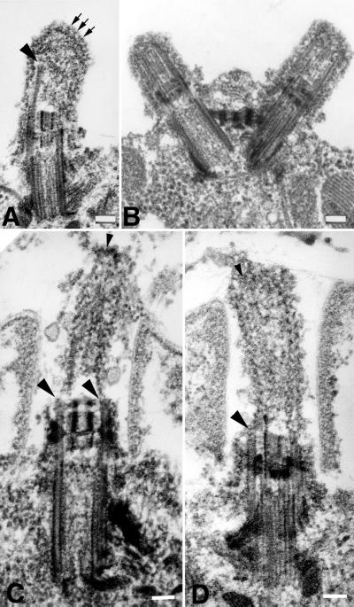
Figure 11.
Electron microscopic analysis of detergent-extracted stf1 mutants. Whole cells were extracted with 0.5% Nonidet P-40 for 5 min (A and B) or 2% Nonidet P-40 for 30 min (C and D) before fixation. Both the cell and flagellar membranes have been removed, but amorphous granular and filamentous material remain associated with the flagellar stumps. Filamentous structures extend from the ends of the flagellar or basal body microtubules (A, C, and D, large arrowheads). In some flagella, the filaments associated with the microtubules coalesce at the distal tip (A, small arrows). In others, microtubules with free proximal ends are occasionally found linked to a distal cap-like structure (C, small arrowhead). Bars, 0.1 μm.

