
Figure 3.
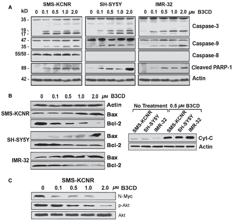
Expression of apoptotic and pro-survival markers in NB cells after B3CD treatment. NB cells (SMS-KCNR, SH-SY5Y, and IMR-32) were treated with increasing concentrations (0.1–2.0 μM) of B3CD for 48 h. Expression of proteins in the lysates of treated and untreated cells by PAGE and Western blot analysis was carried out as described in ‘Materials and Methods’ section. (A) Caspase activation; primary antibodies against pro- and activated caspase-3, -8, -9, and inactivated/cleaved PARP-1. As an internal standard for equal loading, blots were probed with an anti-beta-Actin antibody. (B) Mitochondria-mediated apoptosis; primary antibodies against cytochrome c, Bax, and Bcl-2. (C) Inhibition of N-MYC expression and Akt activation; primary antibodies against N-MYC, Akt, and phosphorylated Akt.