
Fig. 1.
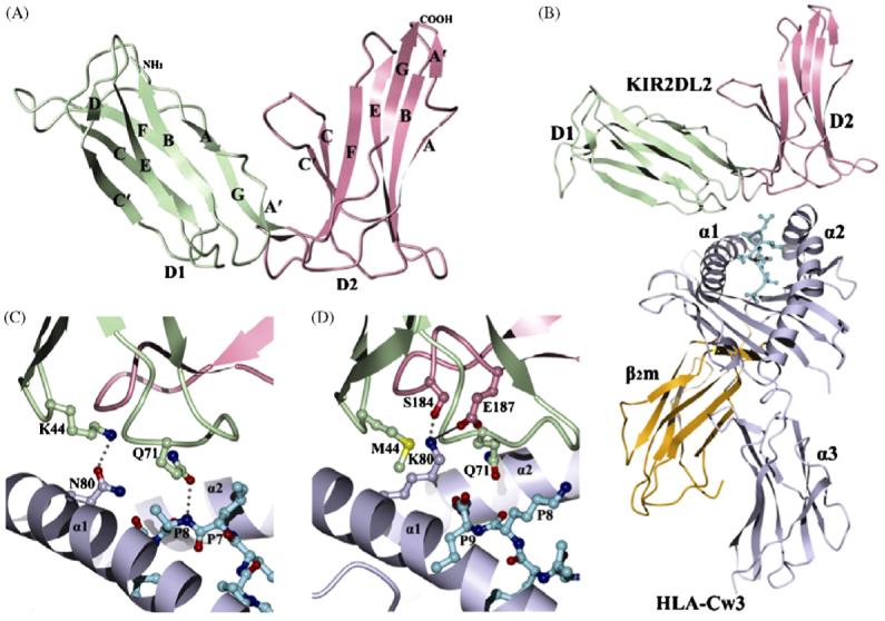
Three-dimensional structures of KIR2DL and KIR2DL/HLA-C complexes. (A) Ribbons diagram of KIR2DL1 (PDB accession code 1NKR). The D1 domain is green; D2 is pink. The secondary structural elements are labeled. (B) Ribbons diagram of KIR2DL2 bound to HLA-Cw3 (PDB accession code 1EFX). The α1, α2 and α3 domains of the HLA-Cw3 heavy chain are blue-grey; β2m is gold; the peptide in ball-and-stick representation is cyan. (C) Cartoon drawing illustrating the allelic specificity and peptide selectivity of KIR2D receptors. The dotted lines represent hydrogen bonds formed by Asn80 of HLA-Cw3 with Lys44 of KIR2DL2, and by Gln71 of HLA-Cw3 with P8 of the peptide. (D) Interactions of Lys80 of HLA-Cw4 (blue-grey) with specificity-determining residues of KIR2DL1 (D1 domain in green, D2 domain in pink) in the KIR2DL1/HLA-Cw4 complex (PDB accession code 1IM9). The solid line represents a salt bridge.