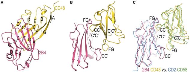
Figure 2. Structure of the 2B4-CD48 Complex.
(A) Ribbon diagram of the 2B4-CD48 complex showing the face-to-face interaction between the AGFCC’C” β sheets of the two IgV domains. 2B4 is rose and CD48 is gold; β strands are labeled.
(B) The complex rotated 90° clockwise about the vertical axis with respect to the view in (A). Loops that contribute to the binding interface are labeled.
(C) Superposition of the 2B4-CD48 and CD2-CD58 complexes (tube diagram). Equivalent Cα atoms of 2B4 were superposed onto CD2, and equivalent Cα atoms of CD48 were superposed onto CD58, with the program Superpose Molecules (Collaborative Computational Project, Number 4, 1994). 2B4 is rose, CD48 is gold, CD2 is cyan, and CD58 is green. The complexes are oriented as in (B).

