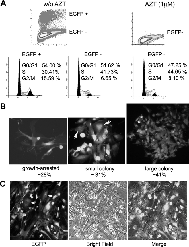
FIG. 5.
HTLV-1-infected-HOS cells escape G1 arrest. (A) HOS/18x21-EGFP cells were cocultivated with MT2 cells with or without (w/o) the addition of AZT as described in the legend to Fig. 3. Three days postinfection, infected and uninfected HOS cells were analyzed by flow cytometry as described in the legend to Fig. 3A for cell cycle profile. EGFP +, EGFP positive; EGFP −, EGFP negative. (B) HOS/18x21-EGFP cells were infected with HTLV-1 as described above for panel A. Cells were trypsinized and plated, grown for 6 days, photographed, and analyzed as described in the legend to Fig. 3B and 4B. The colonies/cell clusters of different sizes were counted as described in the legend to Fig. 4B. (C) HOS cells persistently infected by HTLV-1 were cloned by using limiting dilutions. A representative clone is shown. Cells with mitotic abnormalities are marked by arrows.

