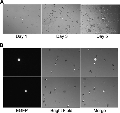
FIG. 7.
HTLV-1-infected Sup-T1 cells ceased proliferation. (A) Half a million 18X21-EGFP SupT1 reporter T cells were cocultured with the same number of MT2 cells in RPMI medium supplemented with 10% fetal bovine serum. After 48 h, cells were collected, counted, dispersed as single cells, and cultured in two 12-well plates at a density of approximately 2,000 cells/well in the same medium. Cells were visualized at 1, 3, or 5 days after limiting dilutions. (B) HTLV-1-infected Sup-T1 cells (EGFP positive) are enlarged and expressed hair-like surface protrusions. EGFP fluorescence and bright-field images and a merged image of the two (merge) of the same visual field are shown. Two randomly selected visual fields from two separate wells are shown.

