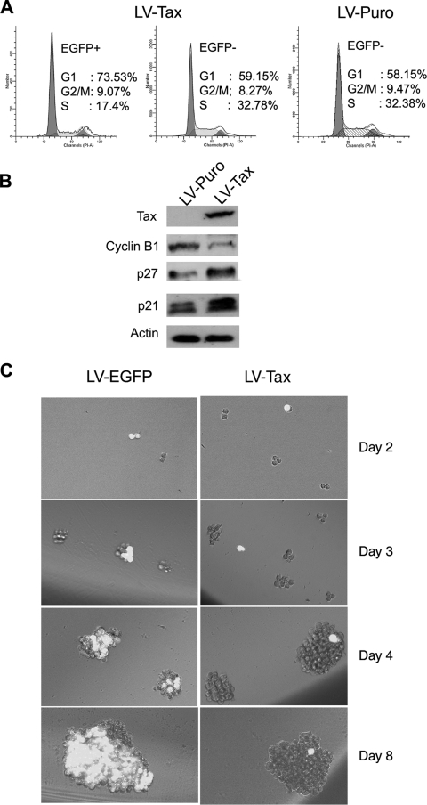
FIG. 8.
LV-Tax-transduced-SupT1 cells ceased proliferation. (A) SupT1/18x21-EGFP reporter cells were transduced with the LV-Tax-SV-Puro or the LV-SV-Puro and cultured for 48 h in RPMI medium supplemented with 10% fetal bovine serum. Cells were collected, fixed, stained, and analyzed by flow cytometry as described in the legend to Fig. 3A. EGFP+, EGFP positive; EGFP−, EGFP negative. (B) SupT1/18x21-EGFP reporter cells were infected with the LV-Tax-SV-Puro or the LV-SV-Puro vectors such that more than 50% SupT1 cells became transduced (based on EGFP expression). Two days posttransduction, cells were harvested and analyzed by Western blotting for cyclin B1, p27, p21, and β-actin (protein control) as indicated. (C) SupT1/18x21-EGFP cells were infected with LV-Tax or LV-EGFP control and grown as described above for panel A. After 48 h, cells were collected, counted, and cultured in two 96-well plates at a density of 5 cells/per well by using limiting dilutions. Cell growth was monitored with an Olympus IX81 fluorescence microscope at 2, 3, 4, and 8 days after the limiting dilution.

