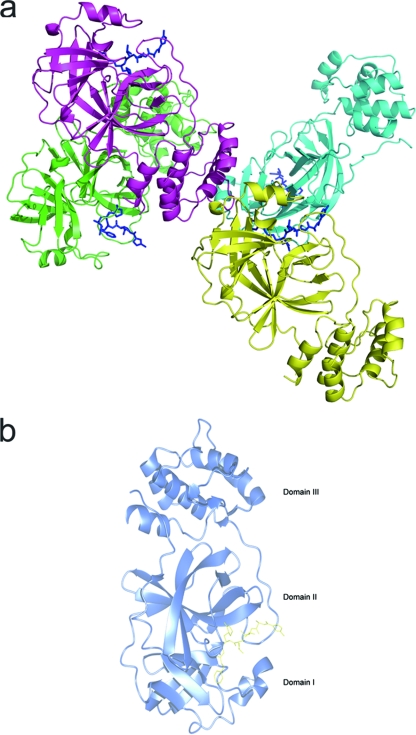
FIG. 1.
(a) Structural overview of four protomers (A, green; B, cyan; C, magenta; and D, yellow) in one asymmetric unit, represented as cartoons. N3 inhibitors are shown as blue sticks. (b) Structural overview of the enzyme-inhibitor complex of one monomer unit. The main chain of the enzyme is represented as blue cartoons, and the synthetic inhibitor is shown as yellow sticks. The three domains are labeled.

