
FIG. 3.
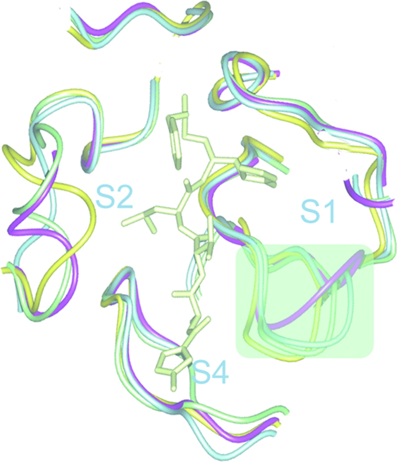
S1 and S2 binding sites of HCoV-HKU1 Mpro main chains of four Mpro structures are superimposed and displayed in the neighborhood of the substrate-binding site. The S1 and S2 binding sites are highlighted by light green shadows. The main chains are represented in worm forms. Different colors are used to represent the strain of CoV. Lemon, synthetic compound; magenta, HCoV-HKU1; light green, SARS-CoV; light blue, TGEV; and yellow, avian IBV.