FIG. 3.
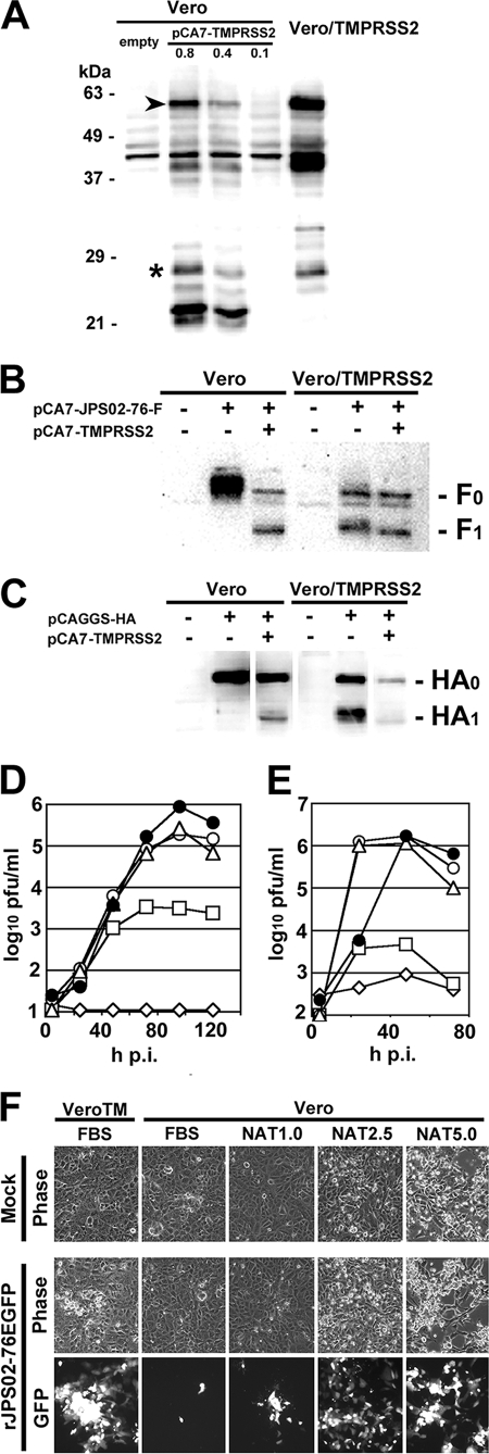
Multiple-step growth of HMPV and influenza virus in Vero and Vero/TMPRSS2 cells. (A) Vero cells were transfected with empty pCA7 vector, or different amounts of pCA7-TMPRSS2 (0.8, 0.4, and 0.1 μg, respectively). TMPRSS2 in these cells at 48 h posttransfection and that in Vero/TMPRSS2 cells were detected by Western blotting. Asterisk, cleaved form; arrowhead, full-length form. (B) Vero or Vero/TMPRSS2 cells were transfected with pCA7-JPS02-76-F alone or together with pCA7-TMPRSS2. At 48 h posttransfection, F0 and F1 proteins were detected by Western blotting. (C) Vero or Vero/TMPRSS2 cells were transfected with pCAGGS-HA alone or together with pCA7-TMPRSS2. At 48 h posttransfection HA0 and HA1 proteins were detected by Western blotting. (D) Vero and Vero/TMPRSS2 cells were infected with rJPS02-76EGFP at a multiplicity of infection of 0.001. Vero/TMPRSS2 cells were cultured in Dulbecco modified Eagle medium (DMEM) supplemented with 7.5% fetal bovine serum (FBS) (•). Vero cells were cultured in DMEM supplemented with 7.5% FBS (⋄) or different concentrations of NAT (1.0, 2.5, and 5.0 μg/ml) (□, ○, and ▵, respectively). Every day after infection, half of the culture media (1 ml) was obtained for plaque titration and replaced with the same amount of respective fresh medium. Plaque assays were performed on Vero/TMPRSS2 cells cultured in DMEM containing 1% methylcellulose and 7.5% FBS. (E) Vero and Vero/TMPRSS2 cells were infected with A/Udorn/72 at an MOI of 0.01. Cells were cultured under conditions as described in panel D, and virus titers in culture medium were determined by plaque assay on MDCK cells. (F) Images of rJPS02-76EGFP-infected Vero/TMPRSS2 (VeroTM) and Vero cells at 2 days p.i. obtained using phase-contrast (phase) and fluorescence (GFP) microscopes. Mock, mock-infected cells; rJPS02-76EGFP, rJPS02-76EGFP-infected cells.

