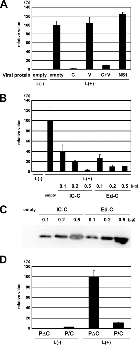
FIG. 2.
Effect of the MV C and V proteins on MV minigenome expression. (A) CHO/hSLAM cells infected with vTF7-3 at an MOI of 0.5 were transfected with p18MGFLuc01, pCAG-T7-IC-N, pCAG-T7-IC-PΔC, and pGEMCR-9301B-L, together with the pCA7 vector expressing the C or V protein of the IC-B strain (C or V), influenza NS1 protein, or empty pCA7 vector. The MV IC-C and IC-V protein-expressing plasmids were also transfected together (C+V). pGEMCR-9301B-L was omitted from a transfection mixture for some cells [L(−)]. At 48 h posttransfection, intracellular luciferase activity was measured, and relative values are indicated. The average value for cells transfected with empty pCA7 vector was set to 100%. The data represent means ± standard deviations of triplicate samples. (B) VV5-4 cells infected with vTF7-3 at an MOI of 0.5 were transfected with p18MGFLuc01, pCAG-T7-IC-N, pCAG-T7-IC-PΔC, and pGEMCR-9301B-L together with various amounts (0.1, 0.2, and 0.5 μg) of pCA7 vector expressing the C protein of the IC-B (IC-C) or Edmonston strain (Ed-C) or empty pCA7 vector. pGEMCR-9301B-L was omitted from a transfection mixture for some cells [L(−)]. At 48 h posttransfection, intracellular luciferase activity was measured, and relative values are indicated. The average value for cells transfected with all three support plasmids and empty pCA7 vector was set to 100%. (C) VV5-4 cells were treated as described in panel B, lysed, and subjected to Western blotting. The C protein of MV was detected using MAb 2D10. (D) CHO/hSLAM cells infected with vTF7-3 at an MOI of 0.5 were transfected with p18MGFLuc01, pCAG-T7-IC-N, and pGEMCR-9301B-L together with pCAG-T7-IC-PΔC or pCAG-T7-IC-P/C. At 48 h posttransfection, intracellular luciferase activity was measured, and relative values are indicated. The average value for cells transfected with pCAG-T7-IC-PΔC was set to 100%. pGEMCR-9301B-L was omitted from a transfection mixture for some cells [L(−)].

